The genetics of porphyria

Image: Pixabay

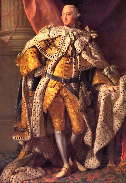
As we’ve already discussed the genetics of one disease back in episode 14, I’d like to focus on another in the form of porphyria- most famously suffered from by George III. However, this is not an isolated case within his family. In fact, porphyria can be seen throughout the family across the centuries, as I discovered when reading two papers from 1968 and 1982 which focus on the ancestors and immediate family of the king. So what causes porphyria and how is it passed down?

Porphyria is actually a group of diseases, one of the main symptoms of which is an increased secretion of proteins called porphyrins into the urine of patients. Porphyrins can also build up in the liver, which may lead to impeded liver function and an elevated risk of liver cancer. Alternatively, the nervous system can be impacted, which may lead to attacks and hallucination.

In porphyria patients, there is a mutation for the enzyme which produces haem according to the British Liver Trust. For reasons I’ll discuss in a moment, I think that this may actually be referring to haemoglobin– a molecule inside your red blood cells which binds them and allows them to carry oxygen through the blood. Haemoglobin is one of the derivatives of porphyrins, which I believe may be why porphyrins then build up- after all, if haemoglobin can’t be produced correctly, it seems logical that the products from the previous step should build up. For this reason, I believe that the ‘haem’ in the British Liver Trust article may refer to haemoglobin, as stated in the Encyclopaedia Britannica.

Most types of porphyria are inherited in an autosomal dominant fashion, meaning that only one copy of this allele needs to be inherited for symptoms to manifest- which might explain how it symptoms kept cropping up in the family shown below. However, there are rarer versions which are recessive, meaning that both copies of the gene need to be faulty before symptoms manifest.

So, that’s porphyria. As an interesting aside, there’s a 2011 article in the New Scientist which mentions that one sufferer is likely to have been Vlad Dracula, which may have started the idea that vampires can’t abide sunlight. In cutaneous porphyria, areas of the skin exposed to sunlight can become blistered. Afflicted individuals consequently avoid sunlight due to pain. Moreover, their skin may shrink back around the mouth, leading to the impression of fangs. I’m not going to go into it article here, but it’s certainly interesting to think that a disease suffered from by kings through the ages may have led to modern ideas about vampires.

A Cause of Alzheimer’s? Part 1- Biofilms

Image: Pixabay

(This material connects to what I discussed in episode 1- Biofilms)

Biofilms- aggregates of material in which bacteria are encased and grow– are relevant structures to fields such as medicine and dentistry, as I mentioned in episode 1. However, when I discussed them back in October 2020, I specifically mentioned their relevance to disease. So, today I thought I’d elaborate a bit and discuss a specific example that is at the forefront of modern research. Specifically, we’re going to talk about Alzheimer’s.

This connection between biofilms and Alzheimer’s is made in a 2020 review paper published in the Clinical Microbiology Newsletter. Although there are other ways of phrasing it, I thought it would be easiest to describe the theory in sequence, starting with infection and ending with the symptoms of Alzheimer’s itself.

So, what happens after biofilm-capable bacteria invade the bloodstream? Bacteria in the mouth have the capacity to form biofilms- to the great frustration of dentists everywhere. It is possible that biofilm fragments of around 100 to 1000 cells, complete with extracellular matrix, can become dislodged, enter the bloodstream and become coated with immune system components. Ordinarily, liver cells called ‘Kupffer cells’- which are capable of phagocytosis- are able to deal with these ‘proto-biofilms’ once they reach the liver. However, if a fragment has all the protective abilities of a biofilm, these Kupffer cells might not be able to digest them. In this case, the fragments can end up in the arterial system. As the aorta pumps oxygenated blood from the heart to the rest of the body, you can imagine that this is a problem.

And this is where genetics and other host factors come into the equation. Ideally speaking, the blood-brain barrier (BBB) should not be permeable enough for these fragments to enter into the brain. However, if the immune system of the host is impaired for whatever reason, such as in the case of diabetes or as a result of normal aging, these biofilm fragments might be able to enter the brain.

This becomes a problem if the biofilm is able to establish itself on the tissues of the brain. In order to obtain nutrients and prevent the host healing, the biofilm provokes constant inflammation, as evidenced by two to three orders of magnitude more interleukins and far more neutrophil immune cells than you would expect. In essence, the biofilm hijacks the immune system to create this inflammation.

There are different host reactions to this chronic inflammation, but the most relevant for our purposes is the formation of amyloid- the formation of large aggregates of amyloid protein in the extracellular space. These amyloid plaques are then supposed to be responsible for the so-called ‘tauopathies’ which are classically associated with the pathology of Alzheimer’s.

Now, it’s important to realise that there is no consensus about what causes Alzheimer’s. For all we know, the biofilm-involved mechanism might not be the ultimate cause. As I highlighted with the possibility that amyloid plaques causing tauopathies, all of these factors may even connected. There are two other factors that have been suggested which I am going to cover in separate posts- a microtubule-binding protein known as tau we mentioned just now (discussed in Part 2) and the possible role of microbiome dysbiosis (in Part 3).